
oral exam guide instrument pdf
The Oral Exam Guide Instrument is a comprehensive resource designed to assist dental professionals in conducting thorough and accurate oral examinations. It provides detailed guidelines, visual aids, and essential diagnostic tools, serving as an invaluable reference for both students and practicing dentists.

1.1 What is an Oral Exam Guide Instrument?
The Oral Exam Guide Instrument is a detailed manual or digital tool designed to standardize and enhance the process of conducting oral examinations. It serves as a step-by-step reference, ensuring that dental professionals can systematically assess various aspects of a patient’s oral health. This instrument typically includes checklists, diagnostic criteria, and visual aids to guide practitioners through inspections, measurements, and evaluations. It is particularly useful for dental students and hygienists, providing a structured approach to identifying conditions such as tooth decay, gum disease, and other abnormalities. By following the guide, professionals can ensure consistency and accuracy in their assessments, ultimately improving patient care outcomes. The instrument is often available in PDF format, making it easily accessible and portable for use in clinical settings or educational environments.
1.2 Importance of the Oral Exam Guide in Dental Practice
The Oral Exam Guide Instrument plays a pivotal role in dental practice by standardizing the examination process, ensuring consistency and accuracy in patient assessments. It serves as a valuable tool for dental professionals, enabling them to identify and document oral health issues effectively. By following the guide, practitioners can detect early signs of conditions such as tooth decay, gum disease, and other abnormalities, leading to timely interventions and improved patient outcomes. Additionally, the guide acts as a learning resource for students and new practitioners, helping them master examination techniques and diagnostic skills. Its structured approach minimizes the risk of oversight, ensuring comprehensive care. The availability of the guide in PDF format further enhances its accessibility, making it a indispensable asset in both clinical and educational settings.
Structure of the Oral Exam Guide Instrument
The Oral Exam Guide Instrument is structured to facilitate systematic dental examinations, featuring sections on patient history, clinical assessment, and diagnostic tools for comprehensive patient care.
2.1 Overview of the Instrument’s Layout
The Oral Exam Guide Instrument is organized into clear, user-friendly sections, ensuring efficient navigation. It begins with patient history and clinical assessment, followed by detailed diagnostic tools and visual aids. Each section is designed to guide professionals through systematic examinations, emphasizing thoroughness and accuracy. The layout prioritizes clarity, with subsections dedicated to specific aspects of oral health, such as periodontal evaluation and restorative assessments. Visual aids, including diagrams and images, complement the text, enhancing understanding and application. This structured approach makes the guide accessible to both students and experienced practitioners, providing a comprehensive framework for conducting precise and effective oral examinations. The logical flow ensures that all critical areas are covered, making it an essential resource for dental practice and education.
2.2 Key Sections of the Oral Exam Guide
The Oral Exam Guide Instrument is divided into essential sections that cover all aspects of a comprehensive oral examination. It includes patient history and clinical assessment, followed by detailed diagnostic procedures. A dedicated section focuses on periodontal evaluation, providing methods for measuring pocket depth and assessing gum health. Another section emphasizes restorative assessments, guiding the identification of decay, fillings, and other restorative work. The guide also includes a section on soft tissue examination, highlighting signs of oral pathologies. Visual aids and diagrams are integrated throughout to enhance understanding. Each section is designed to ensure a systematic and thorough approach, making it easier for professionals to identify and document findings accurately. These key sections collectively provide a robust framework for conducting effective oral examinations, catering to both educational and clinical settings.
2.3 Visual Aids and Diagrams in the Guide
The Oral Exam Guide Instrument incorporates a wide range of visual aids and diagrams to enhance understanding and application of oral examination techniques. High-quality anatomical illustrations provide detailed views of dental structures, while step-by-step procedural diagrams guide users through complex assessments. Color-coded charts and images help differentiate between healthy and pathological conditions, such as tooth decay and gum disease. The guide also includes before-and-after images of treatments, offering practical insights into diagnostic and therapeutic outcomes. These visual elements are complemented by 3D models and interactive features in digital versions, allowing users to explore anatomical details from multiple angles. By integrating these tools, the guide ensures that both students and professionals can visualize and apply examination techniques with precision and confidence, making it an indispensable resource for dental education and practice.
Preparation for the Oral Exam
Preparation involves understanding the exam format, practicing with sample questions, and mastering time management. Utilizing visual aids and familiarizing oneself with instruments like air abrasion tools enhances readiness and confidence during the exam.
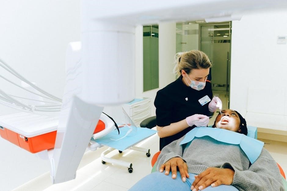
3.1 Understanding the Exam Format
The oral exam format typically includes both clinical and theoretical components, requiring a thorough understanding of diagnostic techniques and patient assessment methods. The clinical portion involves hands-on evaluation, where dentists use instruments like mirrors and probes to examine patients. This section tests practical skills, such as identifying decay or measuring pocket depth, often using tools like air abrasion for precise removal of decayed material.
The theoretical part may include case studies, radiographs, and patient histories, assessing diagnostic accuracy and treatment planning abilities. Familiarizing oneself with the exam structure, including time limits and question types, is crucial for effective preparation. Studying the oral exam guide instrument and practicing with sample questions helps build confidence and ensures readiness for the exam environment.
3.2 Practicing with Sample Questions
Practicing with sample questions is an essential step in preparing for the oral exam, as it helps familiarize oneself with the exam format and content. The oral exam guide instrument often includes realistic scenarios and case studies, allowing candidates to apply their knowledge in practical situations. By reviewing sample questions, individuals can identify areas where they need improvement and refine their diagnostic and treatment planning skills. Timing oneself while answering these questions simulates exam conditions, enhancing time management abilities. Additionally, sample questions cover a wide range of topics, from air abrasion techniques to periodontal assessments, ensuring comprehensive preparation. Regular practice builds confidence and ensures that candidates are well-prepared to address diverse clinical challenges during the actual exam.
3.3 Time Management Strategies
Effective time management is crucial for success in oral exams, as it ensures that all aspects of the examination are thoroughly assessed within the allocated timeframe. The oral exam guide instrument emphasizes the importance of prioritizing tasks, such as starting with critical areas like periodontal probing or decay detection. Allocating specific time slots for each section helps maintain focus and prevents overspending on less critical areas. Practicing under timed conditions during preparation simulates real exam scenarios, allowing individuals to refine their pacing. Additionally, using checklists or mental frameworks can help track progress and ensure no steps are missed. By mastering these strategies, examiners can efficiently conduct comprehensive oral exams while maintaining accuracy and patient comfort.
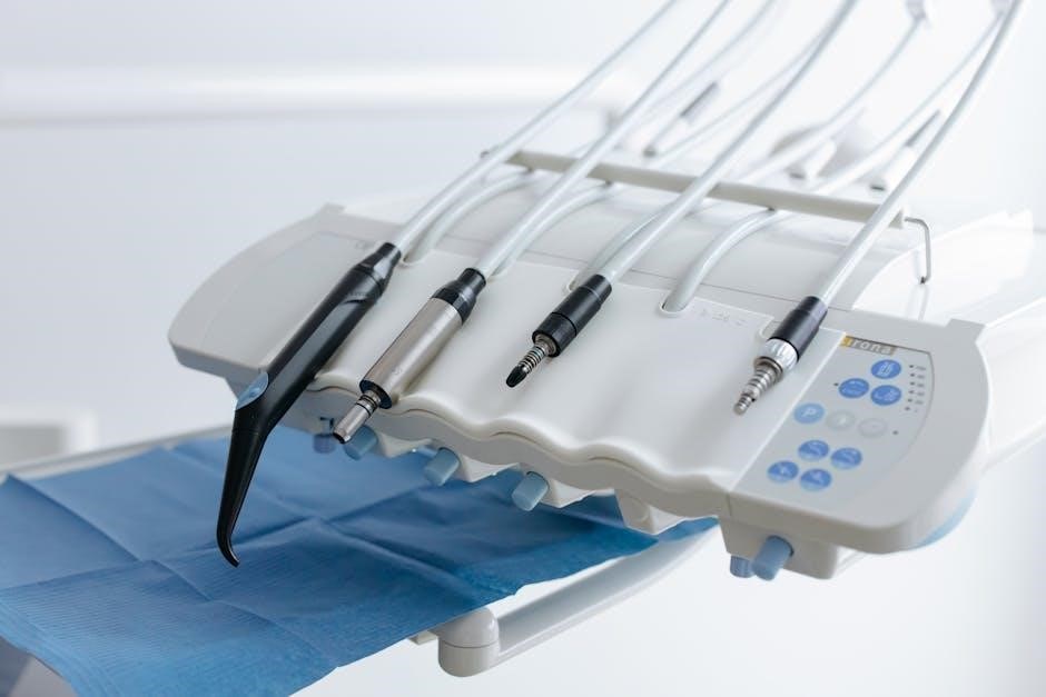
Common Instruments Used in Oral Exams

Common instruments in oral exams include dental mirrors, periodontal probes, and explorers. These tools aid in examining teeth and gums, ensuring accurate diagnoses. They are essential for thorough patient assessments and effective treatment planning.
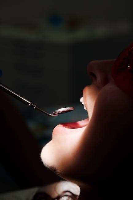
4.1 Dental Mirror: Uses and Techniques
The dental mirror is a fundamental instrument in oral exams, enabling indirect vision and illumination of hard-to-reach areas. It allows clinicians to examine posterior surfaces, detect anomalies, and assess restorations. Proper technique involves holding the mirror at a 45-degree angle and positioning it to reflect light onto the target area. This ensures clear visibility and accurate diagnoses. The mirror also aids in patient communication, as it can display findings. Mastery of mirror handling is essential for effective and efficient oral examinations, reducing patient discomfort and improving assessment accuracy. Regular practice helps refine these skills, making the dental mirror an indispensable tool in daily dental practice.
4.2 Periodontal Probe: Measuring Pocket Depth
The periodontal probe is a crucial instrument for assessing periodontal health by measuring the depth of periodontal pockets. It features graduated markings, typically in millimeters, to ensure precise measurements. Clinicians gently insert the probe into the gingival sulcus, taking care to avoid causing patient discomfort. Pocket depths greater than 3mm may indicate periodontal disease, with deeper measurements often correlating to more severe conditions. Accurate probing is essential for diagnosing gingivitis or periodontitis and for monitoring treatment progress. Regular use of the periodontal probe helps in early detection of periodontal issues, enabling timely interventions. Proper technique ensures reliable results, making the periodontal probe indispensable in routine oral exams for maintaining patient oral health.
4.3 Explorers: Detecting Tooth Decay
Explorers are essential dental instruments used to detect tooth decay during oral exams. These tools feature a pointed or curved tip, allowing clinicians to examine pits, fissures, and surfaces of teeth for signs of demineralization or decay. By gently probing suspicious areas, dentists can identify soft or sticky spots that may indicate early stages of caries. Early detection is critical for preventing progression and minimizing treatment needs. Explorers are particularly useful in areas where visual inspection alone may not suffice. Regular use of explorers in routine exams helps maintain patient oral health by identifying decay early, reducing the risk of more extensive interventions. Their precision makes them invaluable for monitoring high-risk patients and ensuring timely interventions.
4.4 Air Abrasion: An Alternative to Drilling
Air abrasion is a non-invasive technique used in oral exams as an alternative to traditional drilling. It employs a fine stream of particles, similar to a mini sandblaster, to gently remove decayed tooth material or stains. This method is less intimidating for patients, especially children, as it often eliminates the need for anesthesia. Air abrasion preserves more of the tooth structure compared to drilling, making it ideal for early-stage cavities or surface stains. However, it is not suitable for deep cavities or removing old fillings. The process is quiet and reduces the risk of over-preparation, making it a preferred option for minor procedures. By minimizing discomfort and preserving tooth integrity, air abrasion enhances patient comfort while maintaining effective treatment outcomes.
Advanced Techniques in Oral Exams
Advanced techniques in oral exams include air abrasion, laser dentistry, and digital imaging, enhancing accuracy and patient comfort while minimizing invasive procedures and improving overall diagnostic precision effectively.
5.1 Air Abrasion: How It Works
Air abrasion is a modern dental technique that uses a fine stream of particles, similar to a mini sandblaster, to gently remove decayed tooth material or stains. The process involves aiming the instrument at the affected area, allowing the particles to spray and erode the targeted tissue. This method is less invasive than traditional drilling, reducing noise and vibration, which can make it more comfortable for patients. Air abrasion is particularly effective for early-stage cavities and surface stains, as it preserves more of the natural tooth structure. It is also useful for preparing teeth for fillings or sealants. However, it is not suitable for deep cavities or large areas of decay. The procedure is often combined with other diagnostic tools for comprehensive oral exams, making it a valuable addition to modern dental practices.
5.2 Laser Dentistry: Applications in Oral Exams
Laser dentistry has revolutionized oral exams by offering precise and minimally invasive diagnostic and therapeutic applications. Lasers use focused light energy to detect early signs of tooth decay, gum disease, and other oral abnormalities. They are particularly effective in identifying subtle changes in tissue that may not be visible during a routine visual exam. Lasers can also be used to remove plaque, disinfect periodontal pockets, and even treat minor soft tissue lesions during an exam. This technology enhances accuracy, reduces discomfort, and often eliminates the need for anesthesia. Additionally, lasers can aid in detecting oral cancer by identifying abnormal tissue changes. Their versatility and precision make them a valuable tool in modern oral exams, improving both diagnostic capabilities and patient outcomes. As a result, laser dentistry is increasingly integrated into comprehensive oral exam protocols for its efficiency and effectiveness.
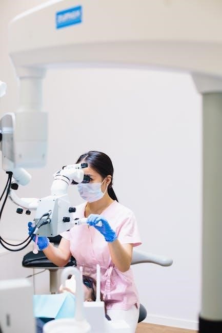
5.3 Digital Imaging: Enhancing Exam Accuracy
Digital imaging has become a cornerstone of modern oral exams, significantly enhancing diagnostic accuracy and patient care. Advanced technologies like intraoral cameras, digital radiography, and 3D cone beam computed tomography (CBCT) provide high-resolution images of teeth, gums, and surrounding structures. These tools enable dentists to detect early signs of decay, fractures, and other abnormalities that may be invisible to the naked eye. Digital images can be enlarged, zoomed, and enhanced, allowing for precise diagnosis and treatment planning. Additionally, digital imaging aids in patient education by visually illustrating oral health issues. The ability to store and compare images over time also supports long-term monitoring and preventive care. By integrating digital imaging into oral exams, dentists can deliver more accurate, efficient, and personalized treatments, ultimately improving patient outcomes and satisfaction.
Tips for Acing the Oral Exam
Effective communication, active listening, and confidence are key to excelling in oral exams. Prepare thoroughly, stay calm, and use visual aids to clarify points, ensuring a polished and professional performance.
6.1 Effective Communication with Patients
Effective communication is crucial for a successful oral exam. Dentists should use clear, simple language to explain procedures, avoiding complex terminology. Active listening ensures patient concerns are addressed, building trust and rapport. Non-verbal cues, such as eye contact and open body language, convey empathy and professionalism. Patients should feel comfortable asking questions, fostering a collaborative environment. Providing reassurance and explaining each step reduces anxiety, making the exam process smoother. Cultural sensitivity and adaptability in communication styles are also essential. Using visual aids from the oral exam guide can help patients understand their conditions better. Regularly checking for understanding and encouraging feedback ensures clear communication. By prioritizing patient-centered dialogue, dentists can enhance the overall exam experience and improve outcomes.
6.2 Maintaining Sterilization and Safety
Maintaining sterilization and safety is paramount during oral exams. All instruments must be thoroughly sterilized using autoclaves or chemical disinfectants before use. Disposable items, like gloves and masks, should be worn to minimize cross-contamination. Surfaces and equipment should be disinfected regularly, following strict infection control protocols. Proper hand hygiene is essential, with handwashing before and after patient interaction. Personal protective equipment (PPE) ensures both patient and clinician safety. The oral exam guide instrument emphasizes adhering to safety standards to prevent the spread of infectious diseases. Regular training on sterilization procedures is recommended to maintain compliance with dental regulations. By prioritizing safety, dental professionals create a secure environment for accurate and effective oral examinations.
6.3 Staying Calm and Focused During the Exam
Staying calm and focused during an oral exam is crucial for accuracy and patient trust. Deep breathing exercises can help reduce anxiety, while positive visualization techniques can build confidence. Proper preparation, including thorough familiarity with the oral exam guide instrument, ensures a sense of readiness. Managing time effectively allows for a systematic approach, preventing rushed decisions. Maintaining eye contact with patients and clear communication fosters a professional environment. Staying present and avoiding distractions ensures attention to detail. Regular practice and mock exams can enhance composure under examination conditions. By combining mental preparation with technical skill, dental professionals can perform at their best, delivering precise and efficient oral exams.

Tools and Resources for Oral Exam Preparation
The oral exam guide instrument PDF offers a comprehensive toolkit, including dental mirrors, periodontal probes, and explorers. It also features study materials, online courses, and practice software for effective preparation.
7.1 Recommended Study Materials
The Oral Exam Guide Instrument PDF is a primary resource for preparation, offering detailed guidelines and visual aids. It is complemented by textbooks on oral pathology and diagnostic techniques, which provide in-depth knowledge. Online study guides and practice exams are also essential, helping candidates familiarize themselves with exam formats. Additionally, flashcards and case studies are valuable for reinforcing key concepts and clinical scenarios. Many dental schools and professional organizations offer supplementary materials, such as video tutorials and interactive modules, to enhance learning. Utilizing these resources ensures a well-rounded preparation, covering both theoretical and practical aspects of oral examinations. By combining the PDF guide with these materials, candidates can build confidence and proficiency in their diagnostic and communication skills.
7.2 Online Courses and Tutorials
Online courses and tutorials are invaluable for mastering the Oral Exam Guide Instrument. Platforms like Coursera and Udemy offer specialized dental examination courses, providing video lessons and interactive modules. These resources often include step-by-step demonstrations of clinical techniques, patient communication strategies, and diagnostic procedures. Many tutorials focus on practical skills, such as using periodontal probes or interpreting radiographs, which are essential for oral exams. Additionally, some courses incorporate real-life case studies, allowing learners to apply their knowledge in simulated scenarios. These online tools are particularly beneficial for self-paced learning, enabling users to review and practice concepts repeatedly. By combining theoretical knowledge with hands-on exercises, online courses enhance proficiency in conducting thorough and accurate oral examinations, making them a crucial supplement to the Oral Exam Guide Instrument PDF.
7.3 Practice Software and Simulators
Practice software and simulators are cutting-edge tools designed to enhance proficiency with the Oral Exam Guide Instrument. These digital platforms offer interactive simulations of oral exams, allowing users to practice diagnosing and treating virtual patients. Many programs include realistic 3D models of teeth and gums, enabling detailed exploration of anatomical structures. Simulators often feature scenarios that mimic real-world clinical challenges, such as detecting decay or assessing periodontal health. Users can practice using virtual instruments, like dental mirrors and periodontal probes, to refine their techniques. Additionally, some software provides instant feedback on performance, highlighting areas for improvement. These tools are particularly useful for students and professionals seeking to sharpen their skills in a risk-free environment. By combining hands-on practice with theoretical knowledge, practice software and simulators complement the Oral Exam Guide Instrument PDF, ensuring a well-rounded educational experience.
The Oral Exam Guide Instrument is an essential tool for dental professionals, offering comprehensive guidance to enhance examination accuracy and patient care. It ensures effective diagnosis and treatment, improving dental practice outcomes.
8.1 Summary of Key Points
The Oral Exam Guide Instrument is a vital resource for dental professionals, providing structured guidance for conducting thorough oral examinations. It covers essential tools, techniques, and best practices, ensuring accurate diagnoses and effective patient care. The guide emphasizes preparation, including understanding exam formats and practicing with sample questions, while highlighting time management strategies for efficiency. It also explores advanced techniques like air abrasion and laser dentistry, alongside the importance of visual aids and diagrams for clarity. Key sections focus on common instruments, such as dental mirrors and periodontal probes, and stress the importance of sterilization and patient communication. By leveraging recommended study materials, online courses, and practice software, professionals can enhance their skills and confidence, ultimately delivering superior dental care outcomes.
8.2 Final Tips for Success
To excel in oral exams, stay calm and composed, as confidence enhances performance. Practice effective communication with patients to build trust and ensure clear understanding. Regularly review the Oral Exam Guide Instrument to reinforce knowledge and techniques. Stay updated with advanced tools like air abrasion, which uses a fine particle stream to remove decay, offering a modern approach to dental care. Prioritize sterilization and safety protocols to maintain a professional and hygienic environment. Allocate time for self-assessment and seek feedback to identify areas for improvement. Utilize recommended study materials and practice software to refine skills. By combining thorough preparation, attention to detail, and a patient-centered approach, you can achieve success and deliver exceptional dental care outcomes.